

Ultra-fast holographic Doppler imaging of the retina
Ocular diseases represent a major global public health challenge. Assessing retinal health requires reliable and precise measurements of local blood perfusion. Yet, even the most advanced imaging techniques—such as fluorescein or indocyanine green angiography and OCT angiography—remain limited in their ability to characterize blood-flow dynamics across the cardiac cycle. This underscores the need for noninvasive methods capable of accurately estimating quantitative biomarkers such as blood flow, arterial wall stiffness, arterial resistivity index, and blood viscosity in retinal vessels.
Since 2005, the development of ultrafast digital holographic imaging devices for ophthalmology (https://arxiv.org/abs/2403.08849) has been addressing this need. These technologies, deployed in clinical research at the Quinze-Vingts Hospital and Rothschild Foundation Hospital (Paris) since 2017, are now being transferred to industry (Lumibird Quantel Medical, EssilorLuxottica) and to numerous academic teams worldwide. A first functional instrument was replicated abroad at the University of Pittsburgh Medical Center (UPMC, USA) in 2022.
Between 2017 and 2022, near-infrared laser holographic imaging achieved key milestones for clinical use. Compliance with laser exposure limits (ISO 15004-2, ANSI Z136.1) was optimized through an innovative diffuse illumination design, enabling wide-field retinal imaging. Doppler holography relies on coherent axial interferometric detection of backscattered light from the eye, followed by digital filtering via singular value decomposition to extract an exploitable optical signal and render high-quality retinal Doppler images. In 2022, real-time medical ultrafast Doppler holography reached a world-first performance, with a 512×512-pixel camera acquiring at more than 20,000 frames per second (video demonstration: https://youtu.be/77bc3V6EmpE).
This interdisciplinary field, pioneered in France by the Langevin Institute, combines the engineering of functional optical imaging systems with clinical ophthalmology research. It is supported by an open-source software ecosystem (https://github.com/DigitalHolography) linking universities, industry, and eye clinics into a computational imaging network, marking a major step forward in optical biomedical imaging.
Since 2022, we have been deploying ultrafast retinal Doppler holography as a noninvasive functional reference exam for the microcirculation. Performed in just a few seconds on an undilated eye, it retrieves arterial and venous velocity waveforms that reflect ocular perfusion pressure and provides hemodynamic, mechanical, and rheological metrics unprecedented in the clinic. An open-source software chain enables real-time high-speed acquisition, aberration-corrected reconstruction, automated biomarker extraction, and generation of a standardized clinical report—now forming the basis of ongoing multicenter studies. These standardized metrics support triage, personalized therapy adjustment, and longitudinal monitoring, with the ambition of intervening at the earliest reversible stages of ocular and cardiovascular disease, before structural damage occurs.
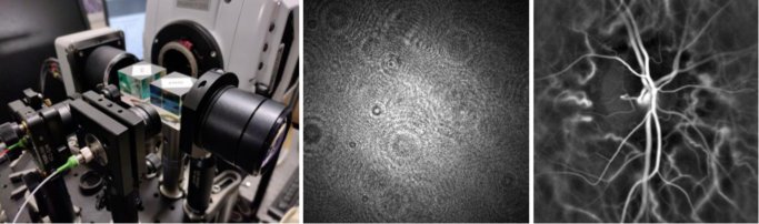
Figure: Clinical holographic Doppler imaging setup (left), recorded interference pattern (center), human retina image (right) reconstructed numerically from the interference patterns.
Numerous internships are available year-round in quantitative analysis of retinal Doppler holography data using Python and MATLAB. Interns will help develop and validate automated pipelines to extract flow-related biomarkers, including blood-flow velocity, vessel-wall dynamics, and rheological metrics. The role involves implementing signal and image processing algorithms, analyzing large datasets, and validating results for clinical applications. This is a unique opportunity to work alongside physicists, engineers, and clinicians at the intersection of computational imaging and medical research. Contact : Michael Atlan.