Homogenized Korteweg–de Vries and Boussinesq models for anisotropic propagation of solitary waves over a structured bathymetry
Pham, K., A. Maurel, and A. Chabchoub
Journal of Fluid Mechanics 1024 (2025)
Résumé: We derive effective Boussinesq and Korteweg–de Vries equations governing nonlinear wave propagation over a structured bathymetry using a three-scale homogenization approach. The model captures the anisotropic effects induced by the bathymetry, leading to significant modifications in soliton dynamics. Homogenized parameters, determined from elementary cell problems, reveal strong directional dependencies in wave speed and dispersion. Our results provide new insights into nonlinear wave propagation in structured shallow-water environments, and consequently motivate further fundamental and applied studies in wave hydrodynamics and coastal engineering.
|


|
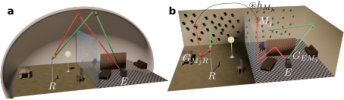
Characterization of temporal aiming for water waves with an anisotropic metabathymetry
Koukouraki, M., P. Petitjeans, A. Maurel, and V. Pagneux
Physical Review B 112, no. 21, 1-9 (2025)

Résumé: The deflection of waves by combining the effects of time modulation with anisotropy has been recently proposed in the context of electromagnetism. In this work, we characterize this phenomenon, called temporal aiming, for water waves using a time-varying metabathymetry. This metabathymetry is composed of thin vertical plates that are periodically arranged at the fluid bottom and which act as an effective anisotropic medium for the surface wave in the long-wavelength approximation. When this plate array is vertically lifted at the fluid bottom at a given time, the medium switches from isotropic to anisotropic, causing a wave packet to scatter in time and deflect from its initial trajectory. Following a simple modeling, we obtain the scattering coefficients of the two waves generated due to the sudden medium change as well as the angle of deviation with respect to the incident angle. We then numerically evaluate this scattering problem with simulations of the full two-dimensional effective anisotropic wave equation, with a time-dependent anisotropy tensor. Finally, we provide experimental evidence of the temporal aiming, using space-time resolved measurement techniques, demonstrating the trajectory shift of a wave packet and measuring its angle of deviation.
|
|
Entropy-controlled velocity-dependent behavior of landslide clayey soil across a wide velocity range
Hu, W., Y. Zheng, Y. Ge, L. Zhou, Y. Li, and X. Jia
Earth and Planetary Science Letters 671, 119671 (2025)
Résumé: The shear resistance of the shear zone governs the behavior of many landslides. Among the factors influencing shear resistance, the velocity dependence of the shear zone can give rise to rapid catastrophic failure (velocity-weakening) or exert a deceleration effect (velocity-strengthening). In this study, we investigated the shear-velocity dependence of shear zone in clayey soil from the Baige landslide in Tibet, China, across a broad range of velocities (3.3 × 10<sup>−8</sup> to 6 m/s), at typical landslide stress levels (200 to 2000 kPa), to simulate the whole lifespan of the landslide, from creep deformation to rapid catastrophic failure. We found that the shear velocity dependence of clayey soils could be classified into three regimes: slight weakening at slow velocities, substantial velocity-strengthening at intermediate velocities, and rapid weakening at high velocities once a critical velocity was attained. The mechanisms for the first and second regimes were explained by the alignment (entropy) change of clay particles along the shear surface. At very slow shear velocities, clay particles become aligned parallel to the shear surface, causing slight weakening and a drop in entropy. As the shear velocity increased, the clay particles became less aligned and more randomly distributed, interlocking with each other, giving rise to strong velocity-strengthening. The rapid weakening in the third regime was associated with frictional heating, independent of entropy. Across the slow to intermediate velocity range, clay particle entropy controls velocity-weakening and velocity-strengthening frictional behavior of shear zones, potentially influencing landslide slow creep. In contrast, rapid shearing causes frictional weakening in clayey shear zones, which may trigger landslide rapid failure. This study offers new insights into landslide dynamics and the transition from creep to rapid failure.
|
|
A diffusion model for light scattering in ejecta
Don Jayamanne, J. A., J. R. Burie, O. Durand, R. Pierrat, and R. Carminati
Journal of Applied Physics 138, no. 20 (2025)
Résumé: We derive a diffusion equation for light scattering from ejecta produced by extreme shocks on metallic samples. This model is easier to handle than a more conventional model based on the Radiative Transfer Equation (RTE) and is a relevant tool to analyze spectrograms obtained from Photon Doppler Velocimetry measurements in the deep multiple scattering regime. We also determine the limits of validity of the diffusive model compared to the RTE, based on a detailed analysis of various ejecta properties in configurations with increasing complexity.
|
|
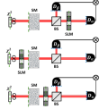
Reconfigurable and active time-reversal metasurface turns walls into sound routers
Dorlot, F., C. Bourdeloux, M. Fink, and F. Lemoult
Communications Physics 8, no. 1 (2025)
Résumé: Sound control in noisy or reverberant spaces is important for applications ranging from communication to immersive audio. However, existing methods often struggle to deliver sound selectively to specific listeners without interference. Here we show that an active acoustic metasurface, composed of programmable elements that both sense and re-emit sound, enables precise targeting of audio in complex environments. Each element processes signals in real time using convolution filtering, allowing us to exploit reciprocity and time-reversal symmetry in wave propagation. Experiments with audible sound in reverberant rooms demonstrate that this approach creates clear, individualized sound channels while suppressing unwanted noise. This research opens new possibilities for adaptive sound delivery in crowded or dynamic settings, with potential applications in conferencing, entertainment, and assistive listening technologies.
|
|
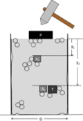
Impulsive shock wave propagation in granular packings under gravity
Van Den Wildenberg, S., X. Nguyen, A. Tourin, and X. Jia
Mechanics Research Communications 150 (2025)
Résumé: We experimentally investigate the impulsive shock propagation caused by an impact into vertically oriented 3D granular packings under gravity. We observe a crossover of wave propagation, from sound excitation at low impact to shock front formation at high impact. One of our findings is a nonlinear acoustic regime prior to the shock regime in which the wave speed decreases with the particle-velocity amplitude due to frictional sliding and rearrangement. Also, we show that the impulsive shock waves at high impact exhibit a characteristic spatial width of approximately 10 particle diameters, regardless of shock amplitude. This finding is similar to that observed in 1D granular chains and appears to be independent of the contact microstructure, whether involving dry or weakly wet glass beads, or sand particles. The final and main finding is that we observe the coexistence of the shock front and the sound waves (ballistic propagation and multiple scattering), separated by a distinct time interval. This delay increases with impact amplitude, due to the increase shock speed on one hand and the decrease of the elastic modulus (and sound speed) in mechanically weakened granular packings by high impact on the other hand. Introducing a small amount of wetting oil into glass bead packings leads to significant viscous dissipation of scattered acoustic waves, while only slightly affecting the shock waves evidenced by a modest increase in shock front width. Our study reveals that shock-induced sound waves and scattering play an important role in shock wave attenuation within a mechanically weakened granular packing by impact. Investigating impact-driven wave propagation through such a medium also offers one way of interrogating a 3D FPUT-like system where nonlinear and linear forces between grains are involved.
|
|


")


.")